ECG Monitoring System
Features
- For each heartbeat, a wave of cardiac muscle contraction pushes blood through the heart.
- Cardiac muscle cells are signaled in succession by an electrical signal that precedes contraction.
- Cells depolarize in a “wave” that causes cells to contract.
- Then the cells repolarize back to normal in succession.
- The signature of the electrical signal as it moves through the heart is measured.
- The ECG is the sum of all depolarization (and repolarization) events.
- The sum of all electrical activity in the heart is illustrated in an ECG trace.
- Display of the signal is a graphical representation of the signal for contraction (and relaxation).
Options
| Order code | System | Size |
| CARDIOPHYS-PRO |
|
19in x 19in x 19in |
| CARDIOPHYS |
|
17in x 13in x 6in |
Benefits
- Produces serviceable ECG signals from small and large animals
- Easy to setup and configure
- Detailed instructions
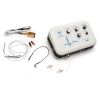
- Package contains the key components
- Amplifier, electrodes, wires
- Exceptional quality components
- High sensitivity
- Low noise
- Reliable source
- >40 years in business measuring small signals
- Personalized customer service (sales and technical)
Applications
- Translational
- Data is similar
- Analysis tools
- Robust analysis of heart function
- Chamber function
- Autonomic control mechanisms
- Channel pathologies
- Physiological phenotype
- Genome-to-phenome (phenomics)
- Toxicants, drugs, etc.
- Stressors (environmental)
- Detailed physiological signatures of effect
- Expansion of lab capabilities
- New (robust) data generation
- Little overhead (i.e. compare to histology, molecular, etc.)
How It Works
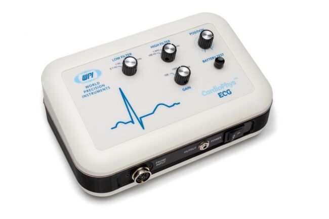
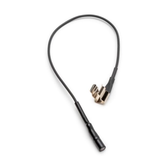
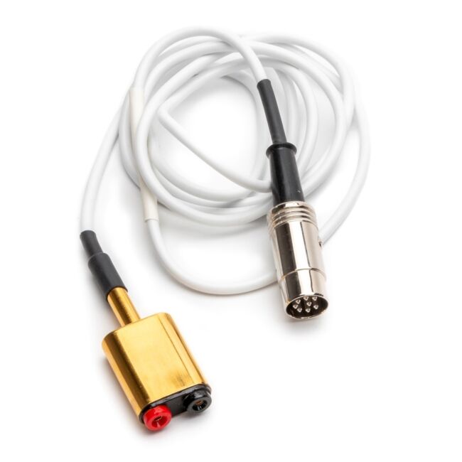
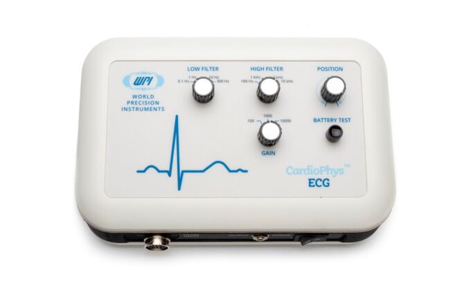
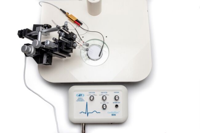
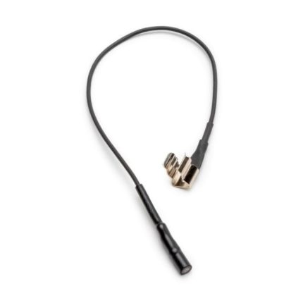
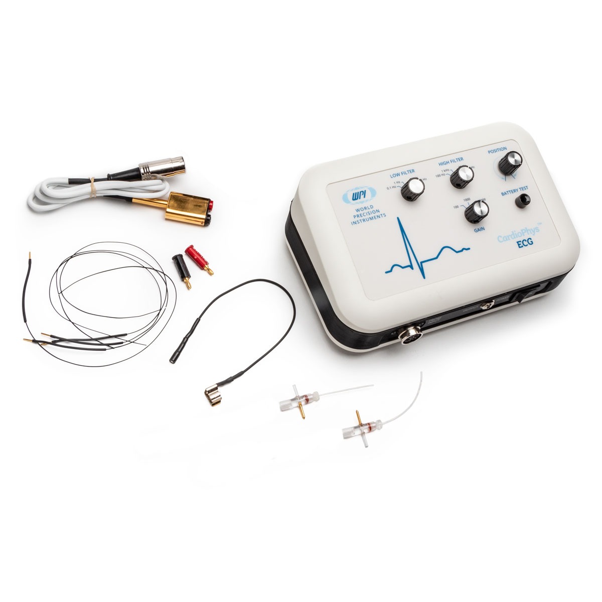
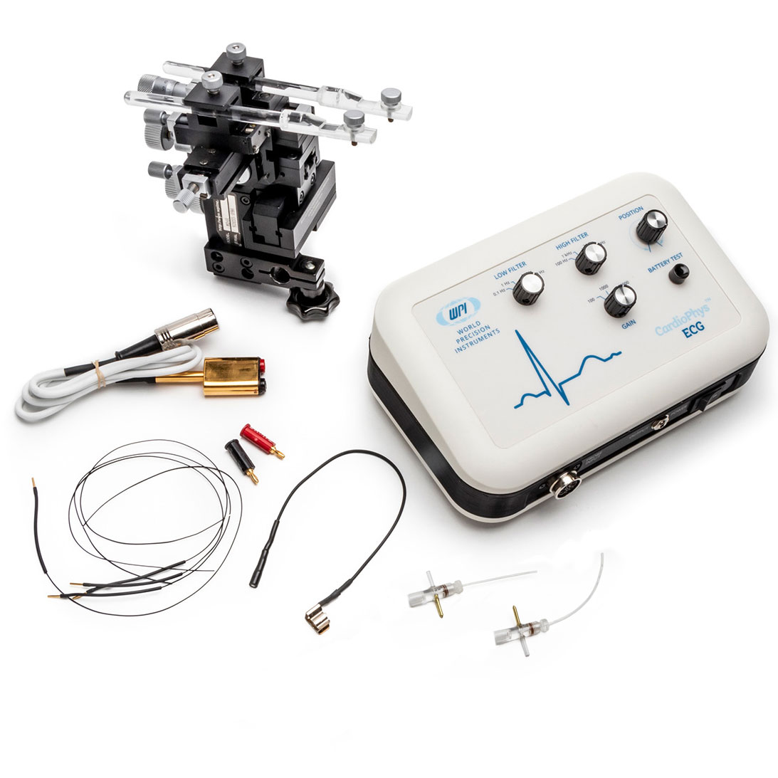
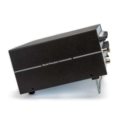
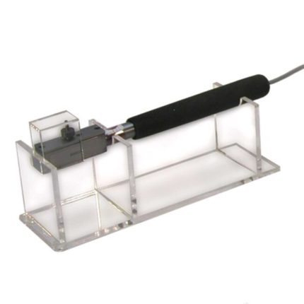
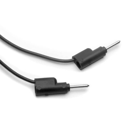
The CardioPhys™ ECG unit receives its input signal from a head stage connected to two electrodes. The electrodes are attached to the animal across the area where an electrical field is generated by the heart. An additional third electrode serves as a ground, which can be either attached to the animal or immersed within a conductive medium surrounding the animal. When the electrodes are attached to the animal, the ground wire serves to complete two circuits, one from each electrode to the common ground electrode. The CardioPhys™ ECG then sends the signal output to a data acquisition system as a voltage signal.
The CardioPhys™ ECG unit has high pass and low pass filters.
- The high pass allows only frequencies above a specified value to “pass,” and eliminates any frequencies below that set value.
- The low pass filter allows frequencies below the specified value to pass and eliminates any frequencies above that set value.
A position dial can adjust the zero, in case of signal drift. The amplification of the signal can be increased from 100X, to 1,000X to 10,000X the incoming signal.
The CardioPhys™ ECG unit includes electronics within the system (both the amplifier and the head stage) that are maximized for low noise recording of very small signals using either glass or metal microelectrodes. The CardioPhys™ ECG unit is versatile enough to measure extracellular voltage signals from animals as small as a hatched zebrafish embryo to animals as large as an alligator (or elephant) to record electrical potentials at the cell or tissue level.
The CardioPhys™ ECG is designed to amplify extracellular biopotentials. These battery powered bioamplifiers incorporate a compact chassis profile that allows the units to be located close to the preparation, which helps minimize long lead lengths that often contribute to noise.
CardioPhys™ Only (CARDIOPHYS)
CardioPhys™ With MD4 Manipulator (CARDIOPHYS-PRO)

